Shane M. Hollawell, DPM reviews his surgical technique for a Lapidus Bunionectomy on a 23-Year Old male utilizing the strong and bio-integrative OSSIOfiber® Trimmable Fixation Nail System.
Case Presentation:
A 23-year-old, 185-pound male presented for initial assessment after a work-related incident. He was suffering from toe pain due to a hyperextension of the right, first metatarsophalangeal joint. The patient noticed immediate drift of his right great toe in the lateral direction. There was no previous medical, surgical or social history relevant to the patient for this case.
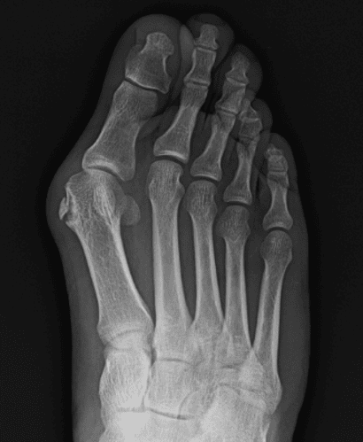
On exam, the patient presented with advanced bunion deformity with prominent medial eminence and increased hallux abductovalgus angle with significant rotation of the hallux. The hallux nail was abutting the second digit. He had joint tenderness on palpation, tenderness with range of motion and general laxity at the first metatarsophalangeal joint. My recommendations at that time remained unchanged: surgically treat the traumatic bunion/turf toe injury, because the deformity worsened, as anticipated. The patient underwent a right foot Lapidus bunionectomy with Treace Lapiplasty® 3D Bunion CorrectionTM, right foot repair of plantar first metatarsophalangeal joint ligament, and right foot excision of fracture fragment from the first metatarsal.
Why was OSSIOfiber® the best choice for this patient?
The OSSIOfiber 2.4 x 30mm Trimmable Fixation Nails provide additional stability required for functional fixation and allow for full bio-integration of the bone-like material properties into the native anatomy of joint surfaces without adverse inflammatory reaction.
Pre-Op Planning:
- In the pre-operative planning stages, the decision was made to perform Lapidus bunionectomy. I utilized 2.4 x 30mm OSSIOfiber Trimmable Fixation Nail as a strong and bio-integrative fixation spanning across the joint surface to assist in the tarsometatarsal joint fusion. The patient underwent a right foot Lapidus bunionectomy, right foot repair of plantar first metatarsophalangeal joint ligament, and right foot excision of fracture fragment from the first metatarsal.
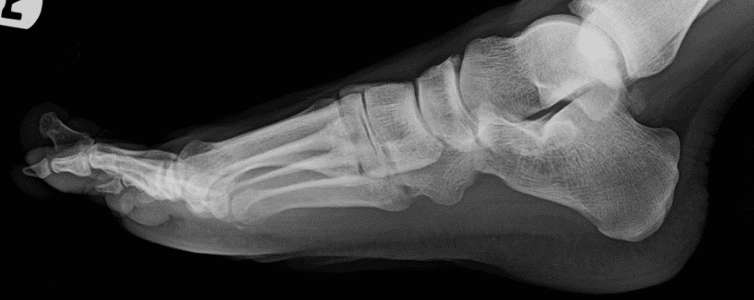
- Traditional X-ray was performed and is shown in the following images:


Surgical Technique with OSSIOfiber Trimmable Nail
The OSSIOfiber 2.4 x 30mm Trimmable Fixation Nail was used in the void left by the removed olive wire fixation.
Incision & Preparation
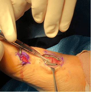
- A #15 blade was brought to the field and used to make an incision overlying the first tarsometatarsal joint. This incision was approximately 3 cm in length.
- The soft tissues were dissected down to the level of the joint capsule. The capsule was incised with 15-blade.
- All soft tissue was reflected off the dorsal, the medial, and the plantar aspect of the joint. The joint was opened with an osteotome.
- Attention was then directed back to the first tarsometatarsal joint, where a Lapidus bunionectomy jig was used to prepare the first tarsometatarsal joint for fusion.
- After bone cuts were made with the sagittal saw, the jig was removed and the cartilaginous fragments were removed from the field.
Primary Hardware Placement
- Both ends of the fusion site were then subchondrally drilled and once completely reduced, provisional fixation with compressive olive wires was placed across the joint.
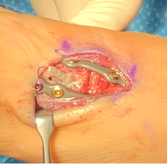
- A Treace Lapiplasty® System 4-hole dorsal locking plate was approximated over the joint and secured in place with four 2.7 mm locking screws.
- A medial locking plate was then approximated across the joint. It was secured in place with four 2.7 mm locking screws.
Insertion of the OSSIOfiber 2.4 x 30mm Trimmable Fixation Nail
- The olive wires used to compress the joint were removed. There was a hole remaining across the joint where the provisional fixation was removed. A tunnel was created with the provided cannulated drill bit to prepare for the insertion of the OSSIOfiber Trimmable Fixation Nail.
- The OSSIOfiber Trimmable Fixation Nail was inserted into the hole utilizing the provided insertion sleeve, tamp and small mallet.


- There were several millimeters of the redundant Nail protruding from the insertion site. This end was trimmed with a bone cutting forceps and re-purposed to fill the deficit left by the second provisional olive wire fixation.


- Deep closure was achieved using 2-0 and 3-0 Vicryl. Skin was reapproximated with 4-0 Monocryl and 4-0 Nylon.

Note: OSSIOfiber material technology has similar radiodensity to cortical bone and is artifact-free on X-ray/CT and MRI safe.
Post-Op Protocol
My post-operative protocol for this patient was as followed:
- Short Cam walking boot and crutches immediately post-operative
- Partial weight-bearing for six weeks
- Normal shoe at 6 weeks
- Full work status at 9 weeks
Summary
The OSSIOfiber® Trimmable Fixation Nail can assist exceptionally in fusion, by providing a solid fixation that supports the locking plate construct, ultimately leaving no permanent implant across the fused joint.
Refer to the product instructions for use for warnings, precautions, indications, contraindications, and complete technique. Medical professionals must use their professional judgement in making any final determinations for product usage and technique.
This case study was created in collaboration with Dr. Hollawell and was developed from his expertise, training, and professional opinion in addition to his knowledge of the OSSIOfiber® Trimmable Fixation Nail. Results from case studies are not predictive of results in other cases. Results may vary.
DOC0001397 Rev 01 12/2020
