Author: Dr. David A. Yeager, DPM at Morrison Community Hospital, in Morrison, IL. Board-Certified Doctor of Podiatric Medicine, practicing both adult and pediatric podiatry.
Education/training: Dr. Yeager DPM, FASPS FACFAS currently practices at Morrison Community Hospital in Morrison, Illinois where he is Chair of Surgery and Chief of Staff. He graduated from Seattle University Honors Program with a dual degree in General Science and English. Dr. Yeager is a doctorate graduate of Rosalind Franklin Chicago Medical School at Finch University where he received his Doctorate in Podiatric Medicine. He completed his residency from Cambridge Health Alliance, affiliated with Harvard Medical School. He is dual Board Certified by the American Board of Podiatric Surgery in Foot and Reconstructive Rearfoot/Ankle Surgery, past Chair of the American Society of Podiatric Surgeons, where he is the current education chair and is a Fellow of the American College of Foot and Ankle Surgeons. He is a past board member of the American Podiatric Medical Association and Past President of the Illinois Podiatric Medical Association. Dr. Yeager also served as residency director and actively promotes educating the young physicians of the profession. He has several publications and was guest Editor for FootandAnkleQuarterly, lecturing both nationally and internationally on a wide range of topics from wound care, advanced foot and ankle reconstruction, to ankle implants.
INTRODUCTION
Enthesopathy describes a disease process specifically involving entheses, the connective tissues between bones and tendons or ligaments. Enthesopathy occurs when these tissues have been damaged, due to overuse, injury or infection. While Enthesopathy may develop in various parts of the body, the entheses of the lower limbs are more frequently involved, and heel enthesitis is the most frequent. Enthesopathy is commonly found in patients over the age of 50 and is usually characterized by symptoms of severe pain and inflammation. Enthesopathy is usually diagnosed by physical examination and a review of symptoms, and in rare cases imaging is necessary. Nonsurgical treatment directed to addressing symptoms is effective for most Achilles enthesopathy cases. Initial treatment consists of ice, activity modification, nonsteroidal anti-inflammatory medications, heel lifts and heel cups, and stretching and strengthening exercises. The final step of conservative therapy is immobilization in a cam walker or cast. Surgery is reserved for the patient who exhausted all nonsurgical treatment options. Surgical approach involves excision of the diseased area of the tendon and calcaneal spur, and reapproximating of the remaining tendon. 1−3
CASE PRESENTATION
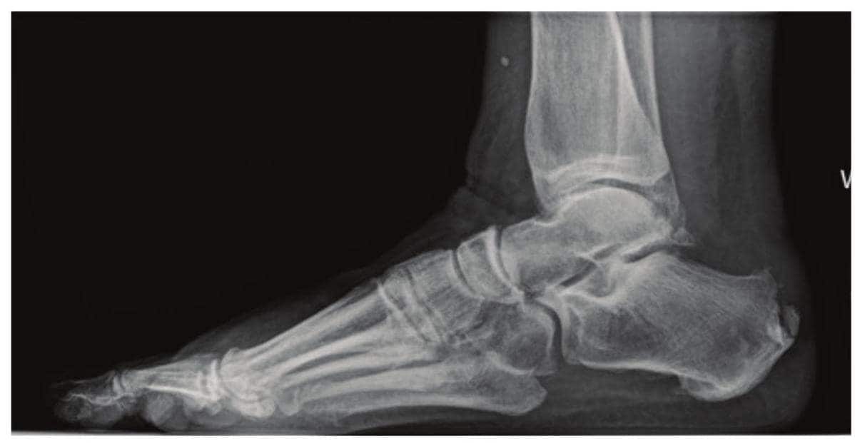
A 58-year-old, female patient, weighting 83Kg presented to the clinic complaining of painful right heel. On examination, her pain was localized to the posterior aspect of the heel area. After unsuccessfully attempting conservative measures including icing, wider shoe gear and Ibuprofen, she opted for surgical correction of the enthesopathy deformity. X-rays were obtained showing substantial posterior heel spur. The patient consented for Achilles tendon lengthening and removal of retrocalcaneal enthesopathy with repair of right Achilles. Patient was made aware of all risks and complications associated with the procedure.
INITIAL ASSESSMENT
Right foot retrocalcaneal enthesopathy with Equinus deformity and Achilles bursitis.
WHY OSSIOfiber IS AN IDEAL CHOICE FOR THIS PATIENT?
The patient was resistant to the use of permanent, not-absorbable fixation techniques due to concerns of potential long-term side effects of metallic hardware and possible need for subsequent removal. She requested OSSIOfiber ® Suture Anchor once it was proposed to her, as it provides fixation stability, without leaving permanent hardware behind.
Preoperative Planning:
Resection of Retrocalcaneal Enthesopathy with Achilles Tendon Lengthening.

Surgical Procedure:
- A V−Y Achilles tendon lengthening, removal of retrocalcaneal enthesopathy heel and Achilles tendon repair utilizing anchor fixation.
Implants Used:
- OSSIOfiber ⊛ Suture Anchor System, 4.75 Anchors w/ Needles ×2
- OSSIOfiber ® Suture Anchor w/ Snare, 4.75 x 1
Surgical Technique Steps:
- Dissection / Access:
Attention is directed to the posterior aspect of the right Achilles area and right heel area where, utilizing a #15 blade, a linear skin incision is performed. This incision is carried down through subcutaneous tissue while protecting and retracting all vital structures. All bleeders are coagulated. Next dissection carried down to the level of the peritenon, which is dissected both medially and laterally exposing the Achilles tendon surgical site. - Tendon Lengthening and Enthesopathy Removal:
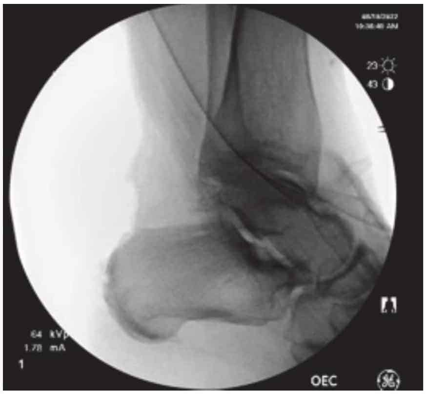
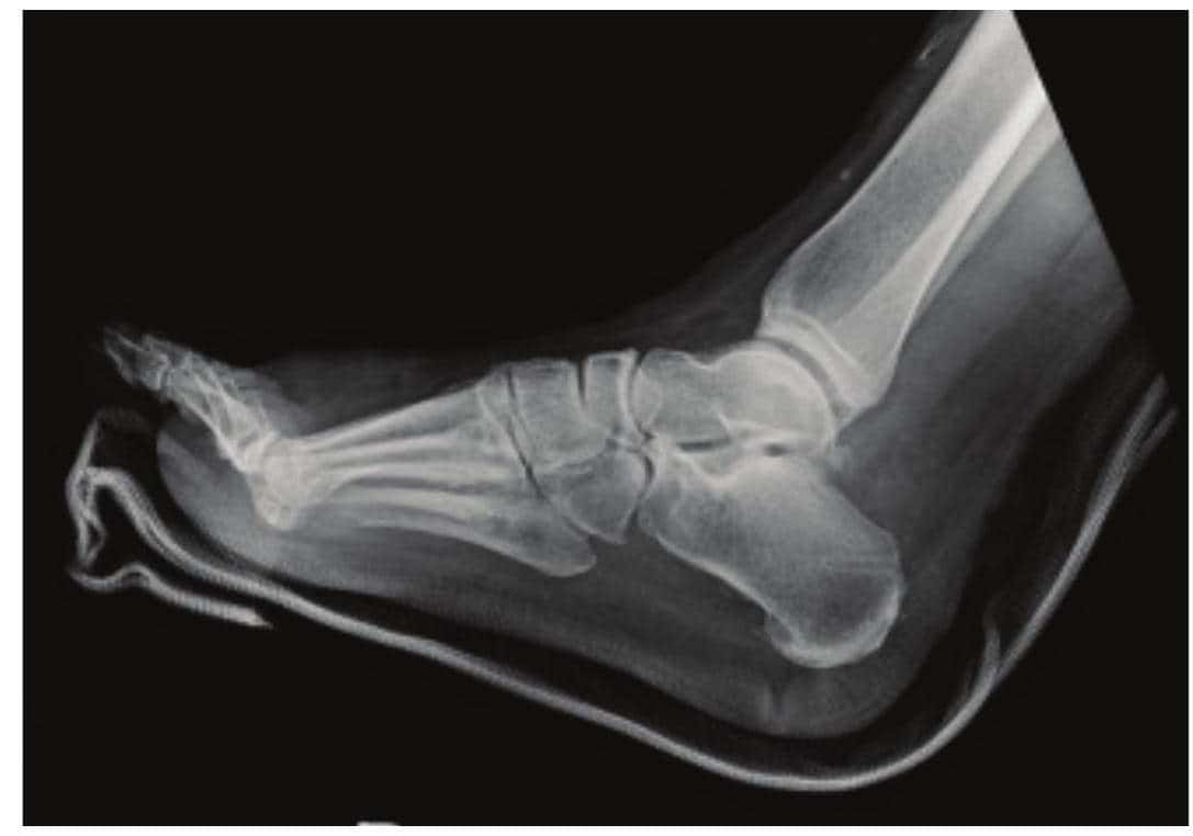
Utilizing a #11 blade, a V cut is made in the central portion of the tendon approximately 7 cm above the superior aspect of the right calcaneus. The tendon is then sutured with 0 Tevdek creating a Y shape. Next, utilizing a #15 blade, a T incision is made over the posterior aspect of the right Achilles area and right heel. This incision is carried down through subcutaneous tissue while protecting and retracting all vital structures. Bleeders are coagulated at this time. Next dissection carried down to the level of the heel spur, noted to be quite substantial. Utilizing a sagittal saw, the superior aspect of the right calcaneus and the posterior heel spur are resected for a more anatomical shape with a smooth surface. This was checked under direct C-arm fluoroscopy and irrigated with 0.9% normal saline impregnated polymyxin solution. - Implants Insertion:
Utilizing 2 OSSIOber ®4.75 preloaded Suture Anchors in the proximal row and one OSSIOber 4.75 snared anchor distally. Three anchors in total were used to not only repair the Achilles but also reapproximate it down to the posterior aspect of the right heel. After the proximal row of anchors were inserted, both sutures were passed through the Achilles Tendon medially and laterally and then hand tied. Taking one of the sutures from the anchor on the medial aspect, the suture was used deep and ran proximally up the tendon and then exited superficially and ran distally down the tendon. Once brought down distally, this suture was then hand tied to one of the sutures from the lateral aspect of the anchor row. The two remaining sutures were then placed through the OSSIOber ⊛ Snared anchor, placed into the most distal hole and tethered down to the bone for close approximation. The foot was then put through a gentle range of motion to ensure the construct is solid. The area was then irrigated. - Closure:
The midline incision through the Achilles tendon is sutured with 2-0 Vicryl, burring the knots in the deep aspect of the tendon. Next, the surgical site is irrigated using 100cc of 0.9% saline. At this time, the peritenon is closed over the tendon using 4-0 Vicryl. The subcutaneous tissue is closed using 3-0 Vicryl, and the skin incision is approximated using 3-0 nylon via horizontal mattress suture technique to prevent skin dehiscence. Approximately 10 mL of 0.5% Marcaine was injected for post-op anesthesia followed by application of wet-to-dry 4×4 s and by dry 4×4 s, Kerlix gauze, Kling roll and a fiberglass posterior mold placed to the patient’s right foot and right lower leg. Just prior to the application of the posterior mold, the tourniquet was released, and immediate warmth and perfusion returned to all digits on the right foot area.
Technique Pearls:
- Tamping the anchors gently before insertion will help the anchor to grip the bone during insertion; there could be an audible squeaking of the anchor while inserting.
- When repairing the Achilles Tendon, it is imperative to go deep first proximally and then superficially for the repair and making sure to connect this strand with one of the suture strands from the other anchor.
- The use of a rasp might be necessary for the contouring of the posterior aspect of the calcaneus.
- Recommend using a drill bit to fenestrate the calcaneus prior to inserting anchors to promote healing.
- When resecting the posterior calcaneal spur, aim the sagittal saw towards the subtalar joint by placing your hand near the subtalar joint as a guide.

Post-Operative Protocol & Patient Follow-Up:
The use of OSSIOfiber ® implants followed the same post-operative weightbearing protocols traditionally utilized for similar cases.
The patient attended scheduled visits up until 2-months post-surgery.
- One week post op – Dressing changed, patient was non-weightbearing with partial cast (posterior mold) applied.
- Week 2 – Sutures removed. At 2 weeks the patient was still non-weightbearing with partial cast applied.
- Week 4 – Transfer to a cam walker boot, still non-weightbearing.
- At 6 weeks the patient was weightbearing in the cam walker boot and began physical therapy.
- At 8 weeks the patient transitioned to a comfort shoe and was fully weightbearing.

Summary:
Bio-integrative technology is specifically beneficial in this case. The patient had astrong objection to metal hardware remaining in her body and OSSIOfiber ⊛ is an all-natural implant, set to gradually bio-integrate in bone, following implantation. The patient’s healing progressed as expected, with an overall favorable clinical outcome, following the use of the OSSIOfiber ® anchors. The quality of the anchors and their overall handling characteristics makes the procedure easy and very reproducible. The OSSIOfiber ® anchor catapults this procedure to a level that each surgeon can attain.
References
1. Alvarez A, Tiu TK. Enthesopathies. [Updated 2023 Jun 5]. In: StatPearls [Internet]. Treasure Island (FL): StatPearls Publishing; 2023 Jan-. Available from: https://www.ncbi.nlm.nih.gov/books/NBK559030/
2. Olivieri I, Barozzi L, Padula A. Enthesiopathy: clinical manifestations, imaging and treatment. Baillieres Clin Rheumatol. 1998 Nov;12(4):665-81. doi: 10.1016/s0950-3579(98)80043-5. PMID: 9928501. Schett G, Lories RJ, D’Agostino MA, Elewaut D, Kirkham B, Soriano ER, McGonagle D. Enthesitis: from pathophysiology to treatment. Nat Rev Rheumatol. 2017 Nov 21;13(12):731-741. [PubMed] [Reference list]
3. Hongli Jiao, E. Xiao, and Dana T. Graves. Diabetes and Its Effect on Bone and Fracture Healing Curr Osteoporos Rep. 2015 Oct; 13(5): 327-335. doi: 10.1007/s11914-015-0286-8
Medical professionals must use their professional judgment for patient selection and appropriate technique.
Results from case studies are not predictive of results in other cases. Results may vary.
Please refer to the product instructions for use for warnings, precautions, indications, contraindications and technique.
Not available for sale outside of the US. Speak to your local sales representative for product availability.
For more information, please visit ossio.io
(®) OSSIO and OSSIOfiber ® are registered trademarks of OSSIO Ltd.
All rights reserved 2023.
DOC-0003109 Rev 1.0
