Author: Cody J. Togher, DPM at The Joint Replacement Institute.
Dr. Togher is a Fellowship-Trained Foot & Ankle Surgeon at the Joint Replacement Institute in Naples, FL. He completed his residency at AdventHealth East Orlando prior to completing fellowship at the Orthopedic Foot & Ankle Center in Columbus, OH. Dr. Togher specializes in complex foot and ankle reconstruction, including total ankle replacement, bunion correction, forefoot and hindfoot deformity correction, sports injuries, and revision surgery.
INTRODUCTION
The naviculocuneiform (NC) joint is located in the middle of the foot and is an integral part of the medial column. It consists of four bones: the tarsal navicular and the medial, intermediate, and lateral cuneiforms. NC joint arthrodesis is used in the treatment of several deformities such as symptomatic fl atfoot and cavovarus foot deformities, posttraumatic arthritis, and degenerative joint disease. The procedure aims to stabilize the medial column and alleviate clinical symptoms. When performing medial column arthrodesis, the NC joint is often a common point of failure due to the mechanical forces acting upon it. Even when performed in isolation, an NC arthrodesis is challenging, especially in the presence of deformity and adjacent joint arthritis. Current literature reports for NC nonunion after attempted arthrodesis is quite diverse, with rates varying from 0 to 24.2%.
CASE PRESENTATION
A female patient presented to clinic 5 months following a previous minimal invasive medial NC joint arthrodesis procedure using 2 metal screws. Patient identified with nonunion confirmed via X-Ray and CT imaging performed within a week of initial presentation. On examination the patient exhibited pain and tenderness with NC passive range of motion, pain with weightbearing, and an inability to perform activities of daily living (ADLs).
WHY OSSIOfiber® IS AN IDEAL CHOICE FOR THIS PATIENT?
Patient believed the metal hardware used in the initial fusion procedure contributed to the nonunion, having reported a history of complications with metallic implants, OSSIOfiber® implants allow good fi xation stability comparable to metal, without the risk for a diffi cult & painful hardware removal process and avoiding permanent hardware remaining in the body.
Preoperative Planning:
Following removal of the existing metal hardware, arthrodesis of all three NC joints and intercuneiform (IC) joints were performed in conjunction with autogenous calcaneal bone graft harvest to promote bone healing of the fusion mass. Autogenous bone graft has demonstrated signifi cant reduction in nonunion rates in midfoot fusion procedures2. Extending the fusion to include the IC joints was thought to increase the available surface area for solid fusion, while also attempting to stabilize the medial cuneiform, in case of continued nonunion. (taking into consideration the history of previous nonunion)
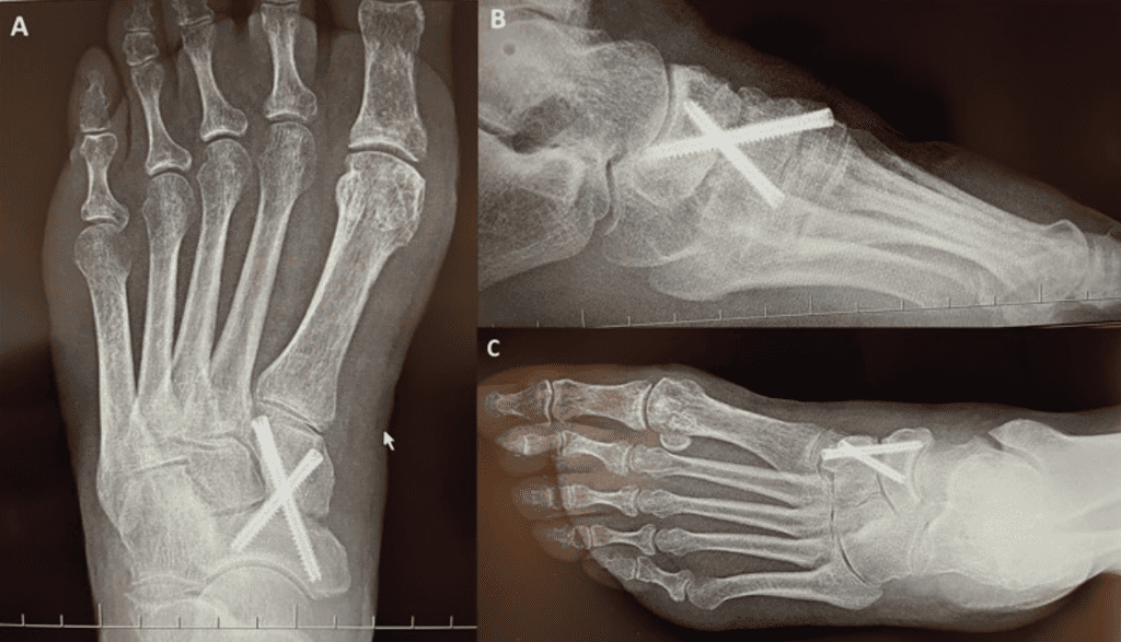
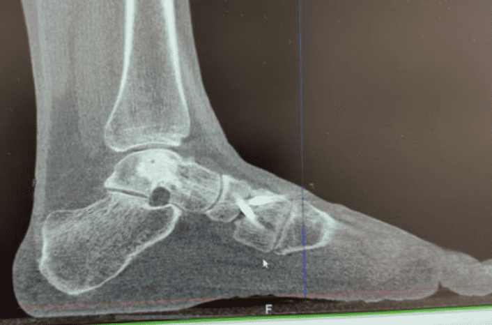
Surgical Procedure:
Midfoot arthrodesis of the NC and IC joints, using an OSSIOfiber® Trimmable Fixation Nail (TFN) and three OSSIOfiber® Compression Screws (CS):
- OSSIOfiber® Cannulated TFN 3.0X50mm
- OSSIOfiber® 4.0 CS (26mm, 38mm & 42mm)
Surgical Technique Steps:
- Dissection / Access:
Dorsomedial approach just lateral to tibialis anterior tendon. Layered dissection with careful retraction of superficial and deep peroneal nerves until subcapsular visualization was achieved. - Fixation site preparation:
Straight osteotomes utilized to break plantar NC ligaments. Hinterman pin distractor was then used to expose joint surfaces which were subsequently debrided using curved osteotomes, curettes, and fenestration drill bit. Cartilage was irrigated from the fusion mass, and bone graft harvested from the calcaneus was then placed into the fusion site. Pin distractors removed. - Tunnel preparation:
The joints were reduced to anatomic alignment, and the fusion mass was temporarily fixated using the wires associated with cannulated screws and TFN. The wires were driven just to the distal cortex and measurements for each corresponding screw and nail were obtained. The wires were then driven further until the apex of the wires were exposed via oscillation on the other side of the foot, where they were secured with a hemostat; this allowed for bicortical drilling without loss of wire purchase/temporary fixation. (Image 3A/B). - Implant insertion:
Once the drill holes were created, the cannulated screws were placed across the three NC joints, followed by nail fixation of the IC joints. - Closure:
Layered closure was performed using #0 and 3-0 Vicryl suture for deep tissues, and 3-0 nylon for skin closure. A well-padded below the knee posterior splint was applied to the operated foot.
Technique Pearls:
- It is important to measure the wire for screw length at the distal cortex, advance the wire, secure with hemostat and then drill for bicortical fixation, avoiding the risk of losing the wire used for cannulated fixation.
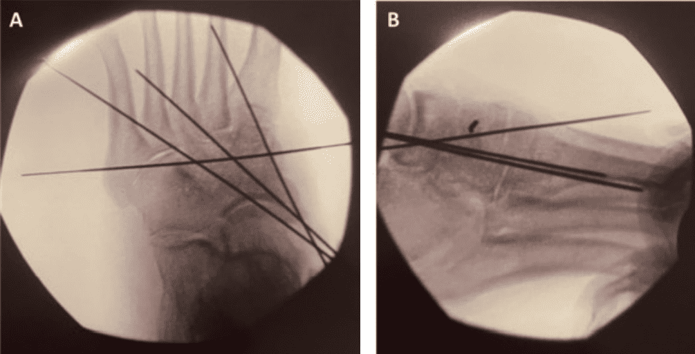
Figure 3: Intraoperative images with OSSIOfiber® fixation implanted. Medial and intermediate screws placed from proximal-medial to distal-lateral; lateral screw placed in distal-lateral to proximal-medial trajection. Nail is placed transversely from medial to lateral.
Post-Operative Protocol & Patient Follow-Up:
Using OSSIOfiber® bio-integrative implants enabled standard post-op protocol. The common post-surgery protocol for this revision procedure is detailed below:
- Non-weight bearing: 8 weeks.
- Partial weight bearing: +4 weeks.
- Full weight bearing: +4 weeks.
- Comfort shoe: same as full weightbearing.
Patient Follow-up:
Patient was able to follow and perform all weightbearing goals postoperatively. CT scan and X-rays obtained at 13 weeks confirmed osseous bridging across the fusion site. Delayed incisional wound healing was noted but completely resolved at 5-6 weeks postoperatively.
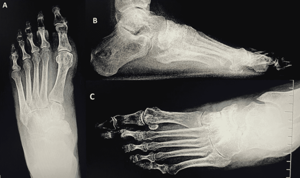
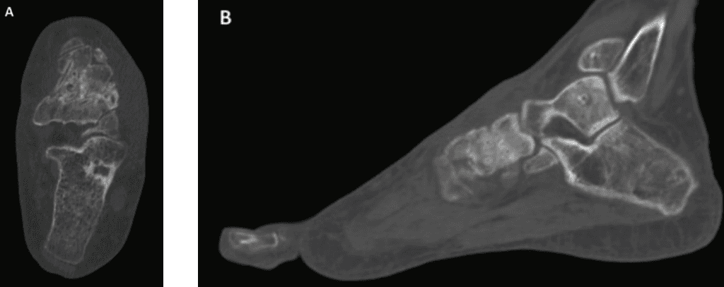
Figure 5: 13 week post-op CT, Axial [A] and Sagittal [B] views, demonstrating joint fusion, bridging across the NC joint. Note OSSIOfiber® fixation allows for better evaluation of the fusion mass and osseous bridging across the arthrodesis without metallic artifacts traditionally observed with metallic fixation.
Summary:
OSSIOfiber® was an exceptional alternative for this patient who suffered symptomatic nonunion and elected for revision but was reluctant to undergo reimplantation of metal. This case demonstrates that together with appropriate joint debridement and preparation, OSSIOfiber® can provide a practical means of fixation; even in a setting where chances of nonunion are elevated.
References
1. Steven A. Tocci, Jacob A. Harder, Andrew M. Ganshirt, David H. Sved, Joseph F. Albers, Daniel W. Guehlstorf, Incidence of Nonunion Following Naviculocuneiform Arthrodesis – A Retrospective Review of 61 Cases, Foot & Ankle Surgery: Techniques, Reports & Cases, Volume 2, Issue 2, 2022, 100196, ISSN 2667-3967, https://doi.org/10.1016/j.fastrc.2022.100196.
2. Buda M, Hagemeijer NC, Kink S, Johnson AH, Guss D, DiGiovanni CW. Effect of Fixation Type and Bone Graft on Tarsometatarsal Fusion. Foot Ankle Int. 2018 Dec;39(12):1394-1402. doi: 10.1177/1071100718793567. Epub 2018 Sep 1. PMID: 30175622. 10.1177/1938640016685153.
Medical professionals must use their professional judgment for patient selection and appropriate technique. Results from case studies are not predictive of results in other cases. Results may vary.
Please refer to the product instructions for use for warnings, precautions, indications, contraindications and technique. Not available for sale outside of the US. Speak to your local sales representative for product availability.
For more information, please visit ossio.io
® OSSIO and OSSIOfiber® are registered trademarks of OSSIO Ltd. All rights reserved 2023.
DOC-0002718 Rev 1.0
