Author: Michael Morwood, MD, Atlantic Orthopaedics, Portsmouth, NH.
INTRODUCTION
The epiphyseal plate (known also as physis or growth plate) is a highly organized cartilage structure between the epiphyseal and metaphyseal bone at the distal ends of long bones. Fractures involving the epiphyseal plate are common musculoskeletal injuries occurring in children with open growth plates. Pediatric ankle fractures represent about 15% of physeal injuries and 5% of all pediatric fractures¹ . Adolescent transitional ankle fractures (ATAF) occur during the closure of the growth phase of the distal tibia when the relatively weak unfused region is prone to fracture. ATAF are more common in children with prior epiphyseal injuries, in adolescents aged 10–15 years, and in males more than females. Sports injuries are the main cause of these fractures2. Since these fractures are accompanied by premature closure of the epiphysis, they may lead to a series of complications such as premature physeal arrest and limb shortening, bar formation, ankle varus/valgus deformities as well as ankle dysfunction. It is necessary, therefore, to achieve accurate anatomical reduction and avoid further damage to the epiphyseal plate during fracture fixation. Although fractures in skeletally immature patients often heal with non-operative management, certain injuries, as those involving growth plates, necessitate operative intervention2,3. Metal implants were traditionally used for fracture fixation, however, there are trending efforts to minimize the presence of permanent implants, mainly in children and adolescents, in active growth4.
CASE PRESENTATION
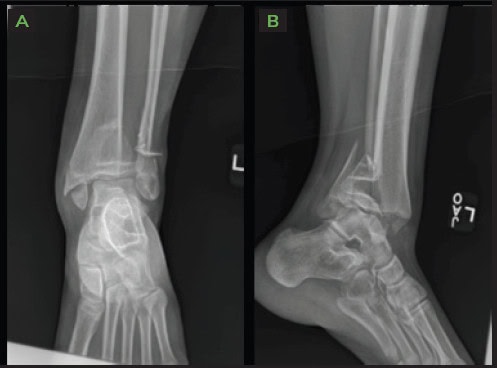
14-year-old female patient who “rolled her ankle” while playing basketball. She is a competitive dancer and is skeletally immature. On examination, she was a mature appearing patient with a swollen, deformed, ankle. Plain film x-rays showed a closed Salter-Harris (SH) type 4 ankle fracture with significant comminution and displacement. The fracture extended through the physis, which is partially fused (consistent with her physiological age), as well as the epiphysis..
The following were noted on x-ray:
- Large metaphyseal fragment with apex anterior fracture angulation (SH-2 portion).
- Comminuted medial malleolar fracture (SH-4 portion).
- Sagittal split in the epiphysis.
Pediatric patients often require metal hardware removal to prevent boney overgrowth and incarcerated hardware later in life. Headless compression screws may be considered to avoid surface hardware/irritation, but would likely become significantly overgrown and incarcerated.
Bio-integrative OSSIOfiber® Compression Screws and Nails provide appropriate fixation stability comparable to metal, while safely eliminating over time. This is the optimal choice for this young patient since it avoids permanent hardware retention in the body, hardware incarceration and secondary removal surgeries.
Pre-operative Planning
Direct anterior approach to the ankle to evaluate and anatomically reduce the joint line as well as the growth plate and medial malleolus.

Surgical Procedure
Open reduction internal fixation (ORIF) of complex distal tibia fracture and fibular fracture.
The following fixation implants were used:
- OSSIOfiber® Cannulated Trimmable Fixation Nail 3.0x50mm x1 unit
- OSSIOfiber® Compression Screw 4.0x34mm x1 unit
- OSSIOfiber® Compression Screw 4.0x32mm x1 unit
- OSSIOfiber® Compression Screw 3.5x32mm x1 unit
Accompanying devices:
- Fibular fracture treated with 1/3 tubular metal plate and screws.
Surgical Technique Steps
Medial dissection-Tibia Fixation:
- Direct anterior approach. A 5 cm incision was made directly over the tibialis anterior tendon. Tib-Ant tendon sheath was opened in line with the incision. Anterior tibial tendon was retracted medially, neurovascular (NV) bundle and extensor hallucis longus (EHL) tendon retracted laterally. Full thickness subperiosteal dissection was carried out medially and laterally, exposing the growth plate which was partially fused.
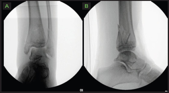
- SH-2 metaphyseal fracture identified and reduced anatomically. Two (2) OSSIOfiber® 4.0mm partially threaded screws placed anterior to posterior.
- Sagittal split in the epiphysis then identified and the joint line reduced anatomically. A single, 3.5mm partially threaded screw placed medially to laterally, all-epiphyseal, parallel to the tibio-talar joint.
- Finally, dissection was carried out medially, identifying a large SH-4 medial malleolus fracture fragment with deltoid ligament attached. This fragment was reduced anatomically and held with bone reduction forceps. A single OSSIOfiber® 3.0mm Cannulated Trimmable Fixation Nail was cut to size and placed. The excess nail was trimmed flush with the bone surface.
Lateral dissection-Fibula Fixation:
- A separate 5 cm incision was made over the fibula fracture site. The subcutaneous border was identified, and full thickness sub-periosteal flaps were developed.
- Fracture manipulated into anatomic reduction and held with bone reduction forceps. 1/3 tubular metal plate of appropriate length applied and affixed with cortical screws, in eccentric mode to further compress the fracture site.
Closure
- Tibiotalar joint capsule closed with 0-vicryl. Anterior tibial tendon sheath closed with 0-vicryl. Skin on both wounds closed with 2-0 monocryl and 3-0 monocryl subcuticular. 1-inch steri strips, xeroform, 4x4s, cast padding and a very well-padded short leg cast was applied.

Technique Pearls
- Place multiple wires at the same time in order to know your trajectories. That way you won’t hit any previously placed screws/nails when multiple fixation points are necessary.
Post-Operative Protocol
- Non-weight bearing: First 6 weeks in short leg cast.
- Transition to partial weight bearing: CAM boot weight bearing as tolerable for about one month, gradual return to normal shoe. Physical therapy for ankle range of motion and strengthening.
- Full weight bearing: by week 10, in a normal shoe.
- Approved to return to sports activities at 3 months.
Patient Follow-up
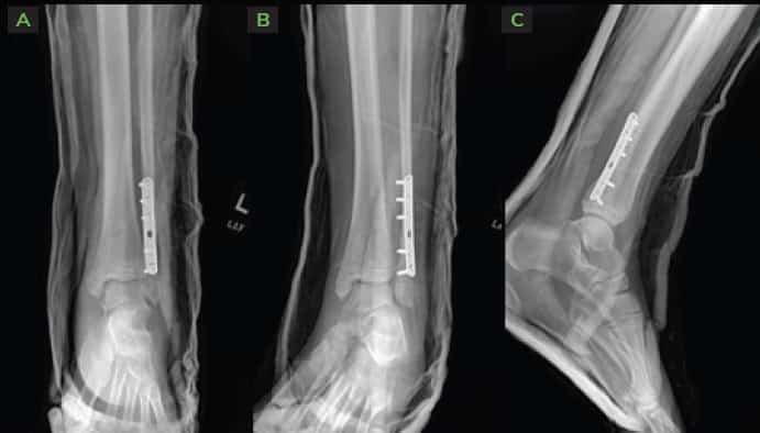
At 2-weeks follow up visit, X-rays confirmed no loss of reduction, all bone fragments well fixated in good anatomical alignment.

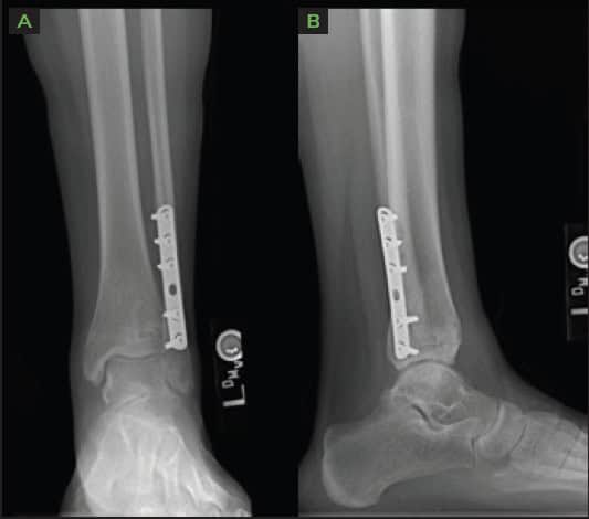
At the 4 months visit X-rays were obtained, demonstrating complete bone fusion of all fracture sites, with excellent joint congruity of all articular surfaces.

Summary:
The OSSIOfiber® implants are a great surgical treatment solution for the skeletally immature population. Utilizing orthopedic permanent hardware in pediatric patients carries more risks due to their longer remaining life span as well as their higher possibility of corrosion, allergic reaction, ion toxicity, malignant transformation, and stress shielding, which are greater in children than in adults4. Instead, the use of bio-integrative implants removes these concerns, as well as avoids boney overgrowth/incarcerated hardware, situations where intercalary or incarcerated segment fixation are needed, i.e. tibial plateau, tibial pilon, and acetabular fixation. Instead of having buried metal screws that will be nearly impossible to remove, this fixation method will completely remodel over time to normal bone, and the need for future removal procedures will be avoided. OSSIOfiber® implants can be incredibly useful also in any other patients with metal allergy concerns.
References
- Venkatadass, K., Sangeet, G., Prasad, V. D. & Rajasekaran, S. Paediatric Ankle Fractures: Guidelines to Management. Indian J. Orthop. 55, 35–46 (2021).
- Pan, Y., Zhang, X., Ouyang, Z., Chen, S. & Lin, R. Transitional ankle fracture management using a new joystick technique. Injury 54, S43–S48 (2023).
- Su, A, W. & Larson, A. N. Pediatric ankle fractures: concepts and treatment principles. Foot Ankle Clin. 20, 705–719 (2015).
- Peterson, H. Metallic implant removal in children. J. Pediatr. Orthop. 25, 107–115 (2005).
