Skip to content
Great news for kids: OSSIOfiber® Metal-Free Compression Screws, Trimmable Fixation Nails, and Compression Staples are now FDA cleared for various bone fractures in children (ages 2-12) and adolescents (ages 12-21). Read More

All-Natural Modified Lapidus for Severe Hallux Valgus Correction 

← All Posts

INTRODUCTION

Hallux Abducto Valgus (HAV) (also referred to as Hallux Valgus (HV) or Bunion) is one of the most common deformities in the foot and ankle field. This pathological condition involves malpositioning of the first metatarsophalangeal (MTP) joint, caused by both a medial deviation of the first metatarsal bone and a lateral deviation and pronation of the great toe. While HAV prevalence reaches 23% in adults 18-65 years of age, it peaks to 35.7% in those older than 65 years. Females have considerably higher prevalence for the deformity1. Additional predisposing factors are family history, metatarsus adductus, first ray hypermobility, pes planus, equinus contracture, ligamentous laxity, and length of the first metatarsal1. HAV symptoms include altered joint mechanics, dysfunction, and progressive pain often at the medial eminence of the first MTP joint. This deformity can vary from mild to severe. More than 100 different operative techniques have been described for the correction of HAV, however no single procedure is universally successful in all patients with severe HAV and even technically well-performed procedures may fail2,3. While mild to moderate HAV can usually be corrected with a distal osteotomy of the first metatarsal, severe HAV deformities with subluxation of the hallux joint, hypermobility of the first tarsometatarsal (TMT) joint, or generalized laxity as well as recurrent HAV following surgical corrections, are the main indications in the literature for a corrective fusion of the first TMT joint, the so-called modified Lapidus. High union rates and high-level of patient satisfaction encourage correction using Lapidus procedure, in particular for severe HAV conditions. 

CASE PRESENTATION

63-year-old, healthy female, non-smoker, with daily routine of moderate physical demands. The patient complained of left foot pain on ambulation, and difficulty with shoe gear. On examination, severe HAV clinical appearance with large deviation of the hallux laterally and hypermobility at the first TMT. The patient requested non-metal surgical fixation. 

Pre-operative Planning

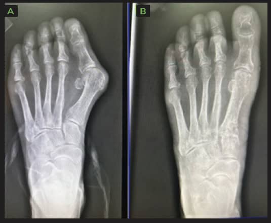
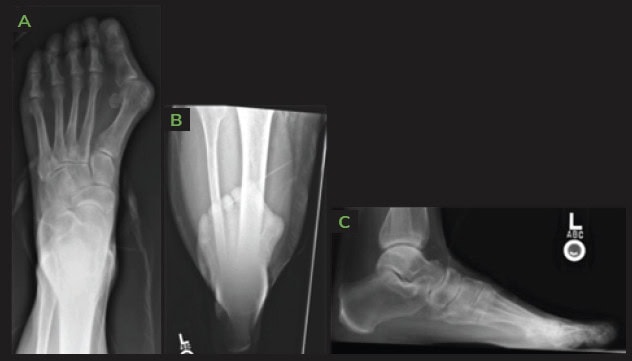
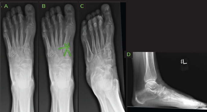
Weight bearing radiographs reveal severe HAV with increase in tibial sesamoid position and 1st Inter Metatarsal (IM) angle. 
Screenshot 2024 09 05 At 18.50.44 Ossio – Naturally Transformative Bone Healing
Figure 1: Pre-operative X-rays; AP [A], AP weightbearing projection [B] and Lateral [C]. 

Surgical Procedure

First TMT joint arthrodesis, Calcaneal autograft harvest. 
The following OSSIOfiber® fixation implants were used: 

  1. OSSIOfiber® Compression Screw 3.5x28mm x 1 unit
  2. OSSIOfiber® Compression Screw 3.5x30mm x 1 unit 
  3. OSSIOfiber® Compression Screw 3.5x32mm x 1 unit 

Surgical Technique Steps

  1. Dissection / Access :
    Dorsal medial 1st TMT incision, lateral release was performed with mini open incision, 1st TMT was hand prepped, and autograft was packed within joint and temporarily fixated with 0.062″ k-wires. A manual reduction was performed. 
  2. Fixation Site Preparation :
    Hand preparation, fenestrated until bleeding bone was present.
  3. Implant Insertion :
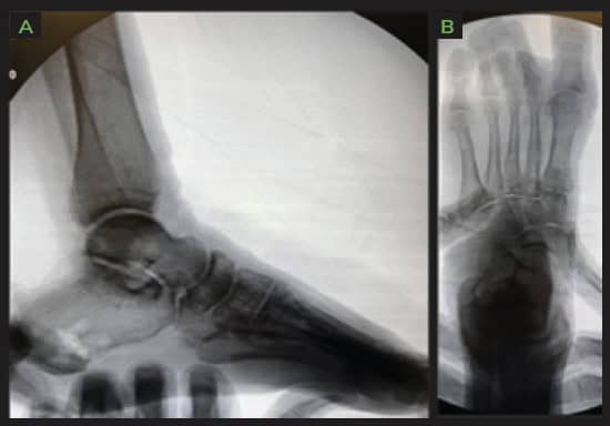
    Three 3.5mm OSSIOfiber® headless Compression Screws were used, two crossing the TMT for compression, third one from M1 to M2 base to hold transverse correction due to large pre op deformity angle.
  4. Closure
    3-0 vicryl for deep and superficial closure, 3-0 nylon for skin. 
Screenshot 2024 09 05 At 19.06.47 Ossio – Naturally Transformative Bone Healing
Figure 2: Intra-operative Fluoroscopy following fixation with OSSIOfiber® devices; AP [A] and Lateral [B]

Post-Operative Protocol

  • Non-weight bearing: 6 weeks 
  • Transition to partial weight bearing: CAM boot for 6 weeks with physical therapy. 
  • Full weight bearing: In supportive tennis shoes after 12 weeks.

Patient Follow-up

The surgical site completely healed by the time the patient came in for the 2 weeks check-up, and sutures were removed. Swelling was minimal over the 1st TMT joint, with minimal discomfort over the joint through initial healing phase. Physical therapy started at 6 weeks post op. 1st MTP joint range of motion improved gradually with passive and active exercises. Sequent radiographs showed well maintained bone reduction and increased signs of fusion from visit to visit. 
Screenshot 2024 09 05 At 19.09.37 Ossio – Naturally Transformative Bone Healing
Figure 3: 6 months Post-operative X-rays: AP [A], AP, illustrated implants demonstrate 3-corner hardware trajectories [B] Oblique [C] and Lateral [D]. 
Screenshot 2024 09 05 At 19.10.17 Ossio – Naturally Transformative Bone Healing
Figure 4: Side-by-side comparison of pre-[A] Vs post- [B] operative X-rays. 
Screenshot 2024 09 05 At 19.11.01 Ossio – Naturally Transformative Bone Healing
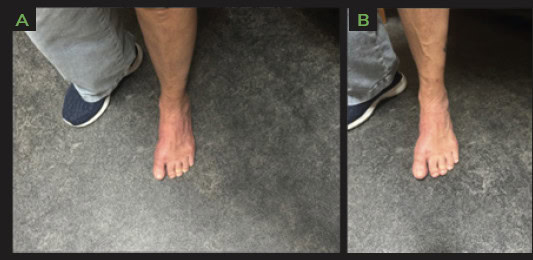
Figure 5: Images of patient’s operated foot 6 months following surgery [A&B]. 

Summary:

First TMT fusions are a reliable and reproducible way to correct severe HAV deformities. Patients are more aware and involved in their medical care than ever before and the trend is moving away from using permanent hardware such as metal/plastics within the body, to a more natural fixation. Bio-integrative implants offer an excellent alternative way of fixating challenging deformities such as the one presented here, providing the required mechanical fixation strength while avoiding the need to deal with retained hardware later in life. 

References

1. Ray, J. J. et al. Hallux Valgus. Foot Ankle Orthop. 4, 1–12 (2019). 

2. Pinney, S., Song, K. R. & Chou, L. B. Surgical treatment of severe hallux valgus: the state of practice among academic foot and ankle surgeons. Foot ankle Int. 27, 1024–1029 (2006). 

3. Fraissler, L., Konrads, C., Hoberg, M., Rudert, M. & Walcher, M. Treatment of hallux valgus deformity. EFORT Open Rev. 1, 295–302 (2016). 

Garret Strand, DPM, Shasta Orthopaedics and Sports Medicine, CA.

Dr. Garret Strand is dual board certified in forefoot/midfoot and rearfoot/ankle surgery by the American Board of Foot and Ankle Surgery (ABFAS) and a fellow of the American College of Foot and Ankle Surgeons, holding the FACFAS certification. As a fellow of the high-volume Northern California Reconstructive Foot & Ankle Fellowship at Shasta Orthopaedics, Dr. Strand practices with exposure to foot and ankle trauma and sports medicine. In his present position as the Assistant Director at the center, Dr. Strand also performs total ankle replacements as well as foot deformity corrections.

Related Posts

Operative Fixation of a Transitional Ankle Fracture in a 14-year-Old Competitive Dancer, Using Bio-Integrative Implants

Author: Michael Morwood, MD, Atlantic Orthopaedics, Portsmouth, NH. INTRODUCTION The epiphyseal plate (known also as physis or growth plate) is a highly organized cartilage structure between the epiphyseal and metaphyseal bone at the distal ends of long bones. Fractures involving the epiphyseal plate are common musculoskeletal injuries occurring in children with open growth plates. PediatricContinue reading “Operative Fixation of a Transitional Ankle Fracture in a 14-year-Old Competitive Dancer, Using Bio-Integrative Implants”

Michael Morwood, MD, Atlantic Orthopaedics, Portsmouth, NH. June 20, 2024

All-Natural OSSIOfiber® Flatfoot Reconstruction

Author: Dallin Greene, DPM, Glendive Medical Center in Glendive, MT. Dr. Dallin Greene is a board-certified Foot and Ankle surgeon, treating all aspects of the foot and ankle with expertise in deformity correction. Dr. Greene graduated from Brigham Young University-Idaho in 2009 with a bachelor’s in biology. He then attended Midwestern University Arizona for aContinue reading “All-Natural OSSIOfiber® Flatfoot Reconstruction”

Dallin Greene April 2, 2024

Midfoot Revision Arthrodesis Using OSSIOfiber® Nail & Screws Following Naviculocuneiform Nonunion

Author: Cody J. Togher, DPM at The Joint Replacement Institute. Dr. Togher is a Fellowship-Trained Foot & Ankle Surgeon at the Joint Replacement Institute in Naples, FL. He completed his residency at AdventHealth East Orlando prior to completing fellowship at the Orthopedic Foot & Ankle Center in Columbus, OH. Dr. Togher specializes in complex footContinue reading “Midfoot Revision Arthrodesis Using OSSIOfiber® Nail & Screws Following Naviculocuneiform Nonunion”

Cody Togher, DPM January 29, 2024

Interested in OSSIOfiber®?

Complete the form and an OSSIOfiber® Advisor will get in touch.

Customer Service

For product inquiries or purchasing questions contact 833-781-7373 or email info@ossio.io

Address

Commercial Headquarters
OSSIO Inc
300 Tradecenter Drive, Suite 3690
Woburn, MA, 01801

Science & Manufacturing Center

OSSIO Ltd.
8 HaTochen Street
Cesarea, Israel 3079861

OSSIOfiber® has been used by 1500+ surgeons across the US

Footnotes

1. Data on file at OSSIO 2. Clinical study data on file at OSSIO 3. Kaiser, P.B., Watkins, I., Riedel, M. D., Cronin, P., Briceno, J., Kron, J. Y. (2019). Implant Removal Matrix for the Foot and Ankle Orthopaedic Surgeon. Foot & Ankle Specialist, 12(1), 79-97. https://doi.org/10.1177/1938640018791015 4. Pre-clinical animal studies (in-bone implantation of OSSIOfiber® and PLDLA control in rabbit femurs). Data on File at OSSIO. 5. Haddad, S. F., Helm, M. M., Meath, B., Adams, C., Packianathan, N., & Uhl, R. (2019). Exploring the Incidence, Implications, and Relevance of Metal Allergy to Orthopaedic Surgeons. Journal of the American Academy of Orthopaedic Surgeons. Global research & reviews, 3(4), e023. https://doi.org/10.5435/JAAOSGlobal-D-19-00023 6. Pot J, Wensen RV, Olsman J. Hardware Related Pain and Hardware Removal after Open Reduction and Internal Fixation of Ankle Fractures. The Foot and Ankle Online Journal. 2011;4(5). doi:10.3827/ faoj.2011.0405.0001.