Author: Bradley W. Lind, DPM, FACFAS, Ryan Foot and Ankle Specialists-Charlotte, NC.
Dr. Bradley W. Lind is a board-certified surgeon in Charlotte, North Carolina who specializes in complex deformity correction of the foot and ankle. An all-natural approach is provided to his patients, when possible, using biointegrative products. He is passionate about providing his patients the best technologies to help restore their anatomy and return patients quickly to their activities.
INTRODUCTION
Flatfoot deformities are a complex disorder characterized by the decrease of the medial longitudinal arch and a valgus deformity of the hindfoot. Due to overuse, the muscles of the limb and foot tend to fatigue and cramp1,2. Surgery for symptomatic flatfoot deformities has been proven to provide pain relief and to improve patient quality of life and physical activity, especially for the young adult population1,3.
The Medial Displacement Calcaneal Osteotomy (MDCO) corrects the hindfoot valgus alignment, resulting in restoration of the medial arch with a neutral pull of the triceps surae through the ankle. The most common indication for an MDCO is the flexible adult flatfoot4.
CASE HISTORY
A 21-year-old male patient, 6’4”, 250lbs, presented with approximately 5 years of intermittent right foot pain, worsening over the past year. The patient is an employee at a factory and spends time on his feet approximately 8-10 hours per shift with significant lateral transfer of weight. His pain is worse with extended periods of time on his foot. Immobilization, home exercises/ stretching, custom molded orthotics, new shoes and diclofenac had only minimal improvement on his condition. He expects to be in mechanical/welding work most of his life and requires a more functional foot to stand on, to avoid direct impact on his livelihood.
CASE PRESENTATION
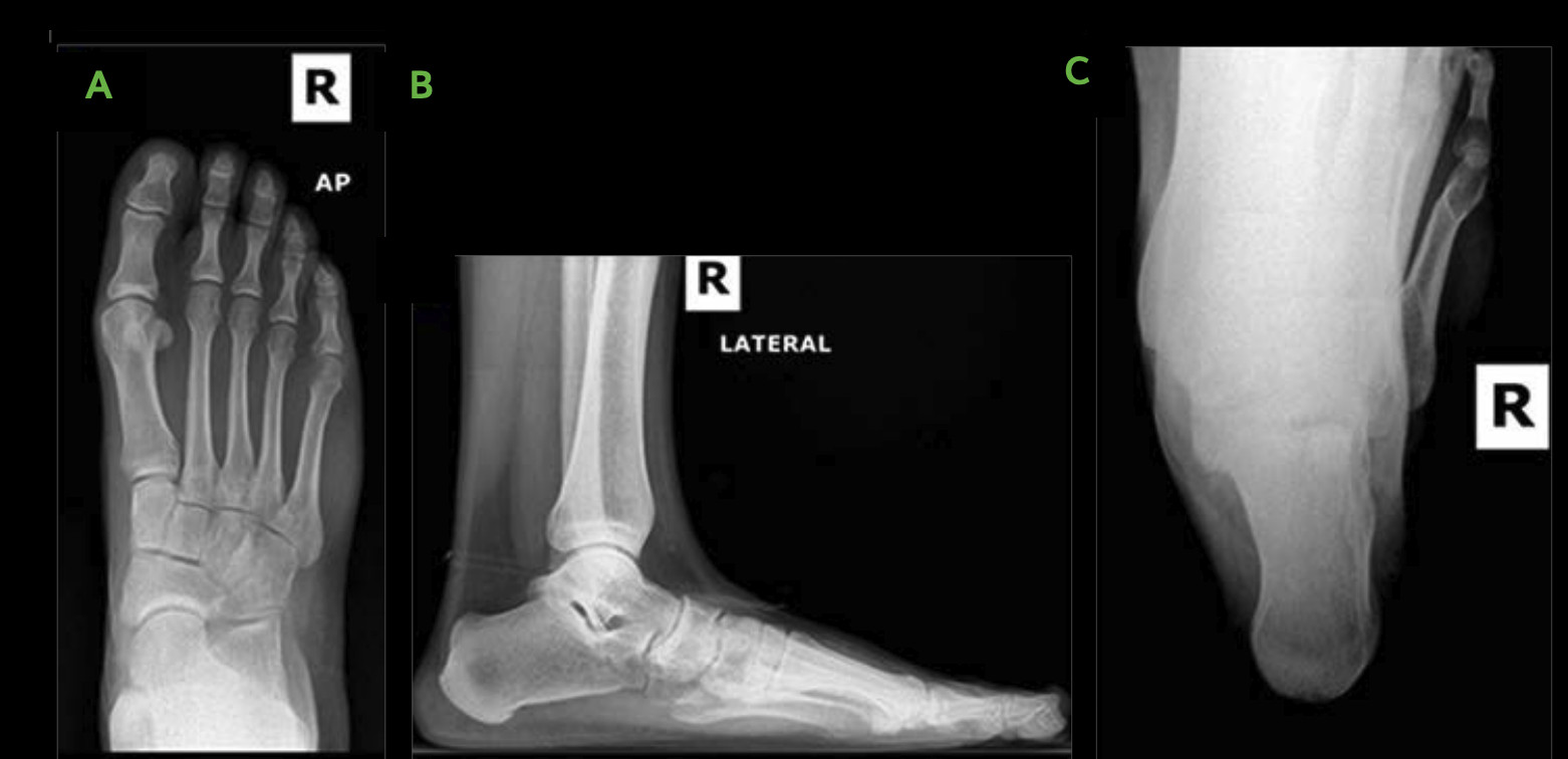
Patient neurovascular status is intact. He presented with overpronation of the midtarsal joint and subtalar joint with diminished height in the medial longitudinal arch, semi-rigid in nature. Hindfoot valgus was noted in neutral calcaneal stance phase with mild resupination on heel rise. Gastrocnemius-soleal equinus demonstrated as well, and no joint crepitus noted on passive range of motion. There is some tenderness along palpation of the posterior tibial (PT) tendon and sinus tarsi and over the lateral cuboid. Mild hallux valgus with hypermobility of the 1st ray and elevatus was noted.
WHY OSSIOfiber® IS AN IDEAL CHOICE FOR THIS PATIENT?
Due to the patient’s young age, the family was interested in a more natural approach for correction. The patient planned to be non-weight bearing approximately 5 weeks, which would allow for moderate consolidation prior to weight bearing.

Surgical Plan
Medial displacement calcaneal osteotomy (MDCO), Evans calcaneal osteotomy, 1st tarsometatarsal joint (TMTJ) arthrodesis with plantarflexion wedge and Akin bunionectomy, gastrocnemius recession, and distal tibial bone graft harvest.
The following OSSIOfiber® fixation implants were used:
- OSSIOfiber® Threaded Trimmable Fixation Nail (TTFN) 5.5x70mm x 3 units
- OSSIOfiber® Cannulated Trimmable Fixation Nail (CTFN) 4.0x70mm x 1 unit
- OSSIOfiber® Compression Screw (CS) 4.0x50mm x 1 unit
- OSSIOfiber® Compression Staple 11x10mm x 1 unit
- MedlineUnite® Evans pre-shaped allograft wedges (6mm and 8mm)
Surgical Technique
The surgeon chose to operate on the patient in two positions.
- Distal tibial bone graft harvest performed first to hydrate both calcaneal and 1st TMT allograft wedges. Next gastrocnemius (Strayer) procedure performed.
- Evans and MDCO in two separate incisions:
For open Evans MDCO procedures Evan’s graft fixated with 4.0mm TFN to avoid excessive compression of allograft wedge and possible loss of correction.
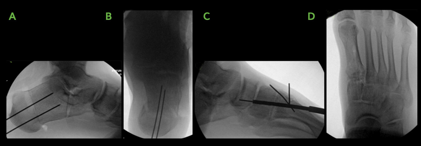
Percutaneous placement of guide wires to fixate MDCO for two parallel OSSIOfiber® 5.5mm TTFNs placed from plantar-posterior heel. - First TMTJ arthrodesis:
Open direct visualization dorsal medial 1st TMTJ, 5.5mm OSSIOfiber® TTFN placed from dorsal central 1st metatarsal into plantar central aspect of medial cuneiform. Additional fixation with OSSIOfiber® CS 4.0x50mm placed from medial 1st metatarsal base into intermediate cuneiform. - Concomitant Akin Bunionectomy:
Small incision lateral to the 1st metatarsophalangeal joint (MTPJ) for lateral release and additional medial incision for the Akin osteotomy, fixated with 11x10mm OSSIOfiber® Compression Staple. - Closure:
Layered closure 3-0 vicryl periosteum and capsule, 4-0 vicryl subcutaneous tissue, 4-0 monocryl running subcuticular suture dorsal incision, 4-0 nylon with horizontal mattress closure to remaining incisions.
Technique Pearls
- No excessive troughing required for Akin small staple
- OSSIOfiber® 5.5X70mm TTFN is an efficient compression device. As with all orthopedic fixation implants, adequate spacing from joint line is required, to avoid stress risers in bone.

showing pilot hole preparation for 1st TMTJ fusion, and AP [D] view, where OSSIOfiber® TTFN & CS implants are visible, crossing from the 1st metatarsal into the medial and intermediate cuneiform bones.
Patient Post-Operative Protocol Follow-up
- Non-weight bearing: 5 weeks
- Partial weight bearing: week 5 to week 6
- Full weight bearing: at 6 weeks
- Back to comfort shoe: at 8 weeks
Patient Follow-up
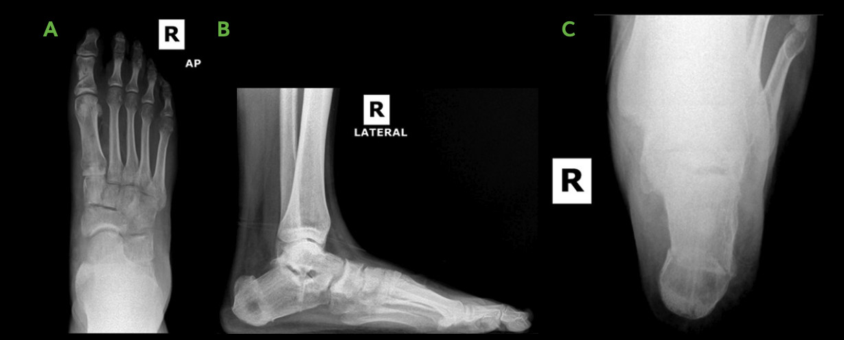
It was easy to visualize the osteotomy/arthrodesis healing progress without metallic hardware in the way, allowing the patient better confidence to start ambulating early. Less hardware related pain was reported by the patient, when compared to metal hardware use in cases of similar nature, especially during wintertime as no metal was present to expand/contract.

Summary:
The all-natural flatfoot repair for lateral column overload performed on this young adult shows the resiliency of the OSSIOfiber® material and implants. Based on personal experience using OSSIOfiber® implants in other similar procedures, the surgeon was confident with allowing this 21-year-old patient to begin bearing weight at 5 weeks. A case of this nature would have previously performed with metallic staples/screws, which only increases likelihood of the patient requiring hardware removal in his or her lifetime due to a young age.
References
1. Martinelli, N., Bianchi, A., Prandoni, L., Maiorano, E. & Sansone, V. Quality of Life in young adults after flatfoot surgery: A casecontrol study. J. Clin. Med. 10, 451 (2021).
2. Lee, M., Vanore, J., Thomas, J. & Catanzariti, A. R. Diagnosis and treatment of adult flatfoot. J. Foot Ankle Surg. 44, 78–113 (2005).
3. Day, J. et al. Outcomes of idiopathic flexible flatfoot deformity reconstruction in the young patient. Foot Ankle Orthop. 5, (2020).
4. Stufkens, S., Knupp, M. & Hintermann, B. Medial displacement calcaneal osteotomy. Tech. Foot Ankle Surg. 8, 85–90 (2009).
DOC-0003993 Rev 1.0 05/2025
Medical professionals must use their professional judgement for patient selection and appropriate technique.
Results from case studies are not predictive of results in other cases. Results may vary.
Refer to the product Instructions for Use for warnings, precautions, indications, contraindications and technique.
Some products mentioned in this document may not be currently available or approved for sale in your country.
Speak to your local sales representative/distributor for product availability.
® OSSIO and OSSIOfiber® are registered trademarks of OSSIO Ltd. All rights reserved 2025.
